Diabetes Awareness Month – Seeing the bigger picture in diabetic eye care
Keeler
November is Diabetes Awareness Month, with World Diabetes Day on 14 November providing an important opportunity to raise awareness of one of the potentially sight-threatening complications of diabetes: diabetic eye disease.
For someone living with diabetes, protecting vision is not simply about having an eye examination. It is about creating an ongoing picture of eye health: detecting retinal changes, understanding what those changes mean, documenting them, deciding whether further investigation or treatment is required and helping patients understand why continued care matters.
At Keeler, we believe ophthalmic technology and clinical education can support both eye care professionals and their patients throughout that journey.
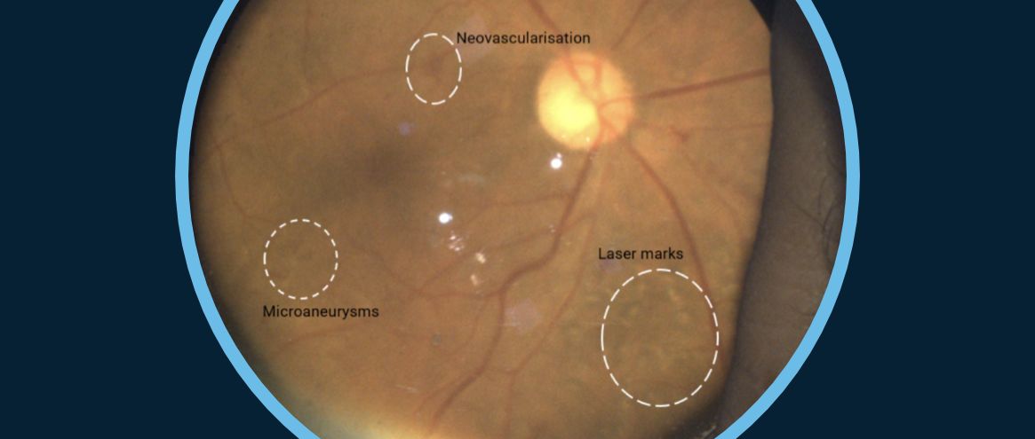
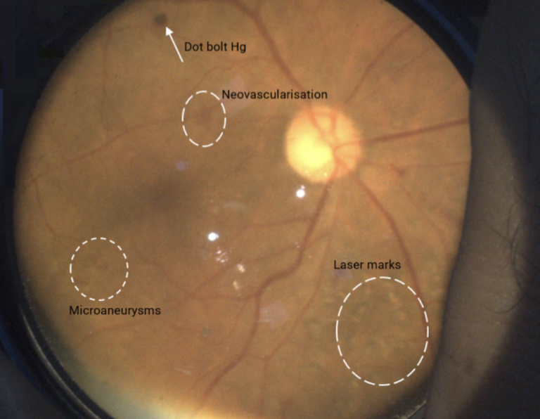
Diabetes can affect the eyes before vision changes
Source: Dr Kenshuk Marwah with patient consent
One of the challenges of diabetic retinopathy is that patients may not experience noticeable visual symptoms during its earlier stages.A patient can therefore feel that their vision is perfectly normal while changes are already occurring within the retina.This makes regular diabetic eye screening and appropriate ophthalmic assessment particularly important.
Retinal examination remains fundamental to understanding diabetic eye disease
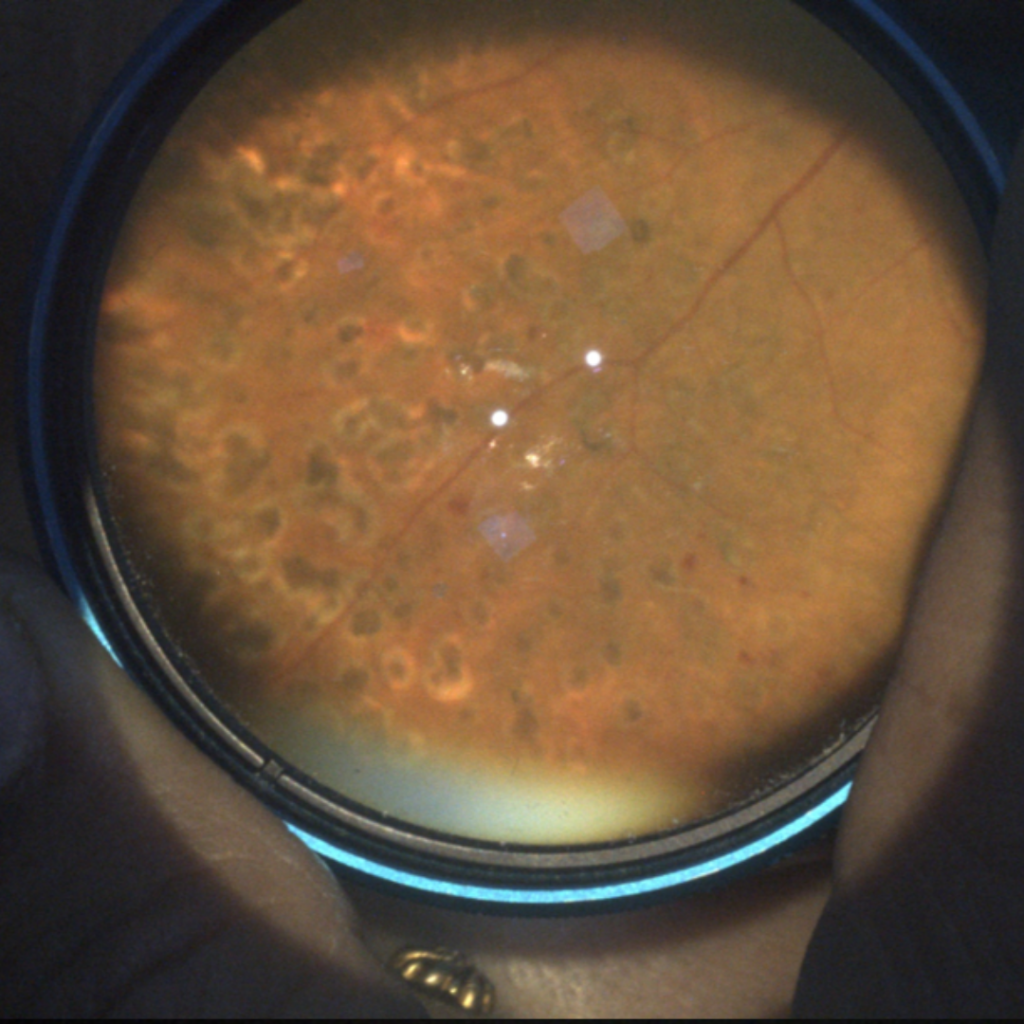
No single examination or imaging modality necessarily provides every answer for patient care in diabetic retinopathy. However, binocular indirect ophthalmoscopy allows the clinician to dynamically examine the retina and navigate towards different areas of interest, including the retinal periphery.
Adding digital capability offers something further: an opportunity to document what is observed. The Keeler Vantage Plus Digital brings digital image and video capture to binocular indirect ophthalmoscopy, allowing findings from the examination to become part of the patient’s visual record.
This can be particularly valuable when assessing the retinal periphery and when more traditional tabletop imaging may be challenging for the patient.
Older or less mobile patients, wheelchair users and some children may find positioning and maintaining fixation at fixed tabletop equipment difficult. Portable examination can also provide greater flexibility in appropriate hospital, community and outreach environments.
Understanding Retina Examination
Source: Anonymous with patient consent.
Source: Dr Kenshuk Marwah with patient consent
The visible retinal examination is only one part of the patients story board. Optical coherence tomography (OCT) provides cross-sectional information about retinal structure and plays an important role when diabetic macular oedema is suspected or requires monitoring. This also provides an important educational opportunity.
A retinal image can help a patient understand where changes are occurring, while an OCT image can help the clinician explain structural changes involving the macula and why these may be important to central vision.
Different technologies and diagnostic equipment therefore contribute different pieces to the same clinical story.
Understanding the retinal circulation with fluorescein angiography
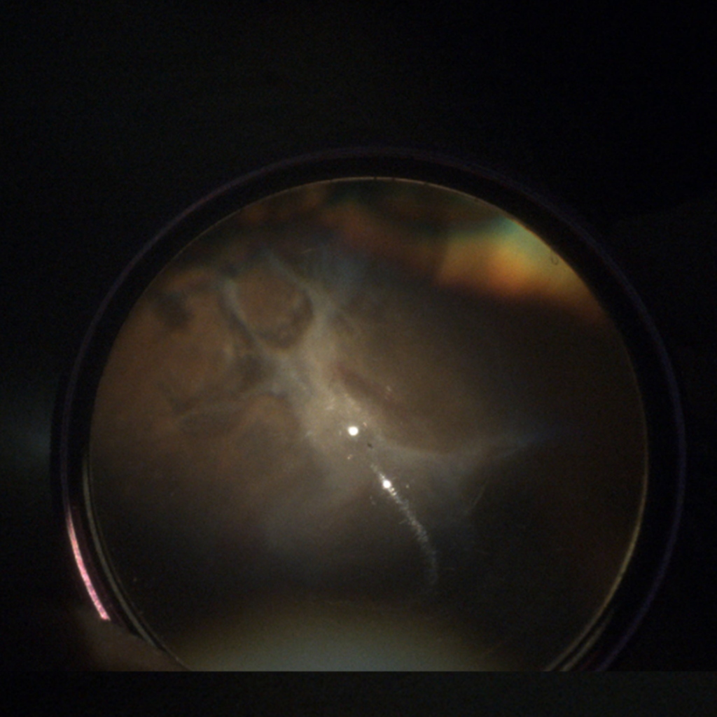
In selected patients, clinicians may need additional information about the retinal circulation.Fluorescein angiography (FA) can provide information about features such as vascular leakage, retinal non-perfusion and neovascularisation.These findings can be particularly relevant in more advanced diabetic retinopathy.
Within an appropriate fluorescein angiography pathway and compatible FA imaging configuration, Digital BIO imaging can provide an opportunity to observe and digitally document retinal fluorescence during a dynamic indirect examination.
This can be especially interesting when the clinical question extends beyond the posterior pole. The clinician can direct the examination towards different areas of the retina, including more peripheral areas, adding another perspective to the patient’s retinal record.
The VPD does not itself perform fluorescein angiography but allows visualisation using injectable flouoresceine. FA remains an invasive diagnostic investigation requiring an appropriate clinical environment, patient consent, appropriately trained staff and established procedures for recognising and managing possible adverse reactions.
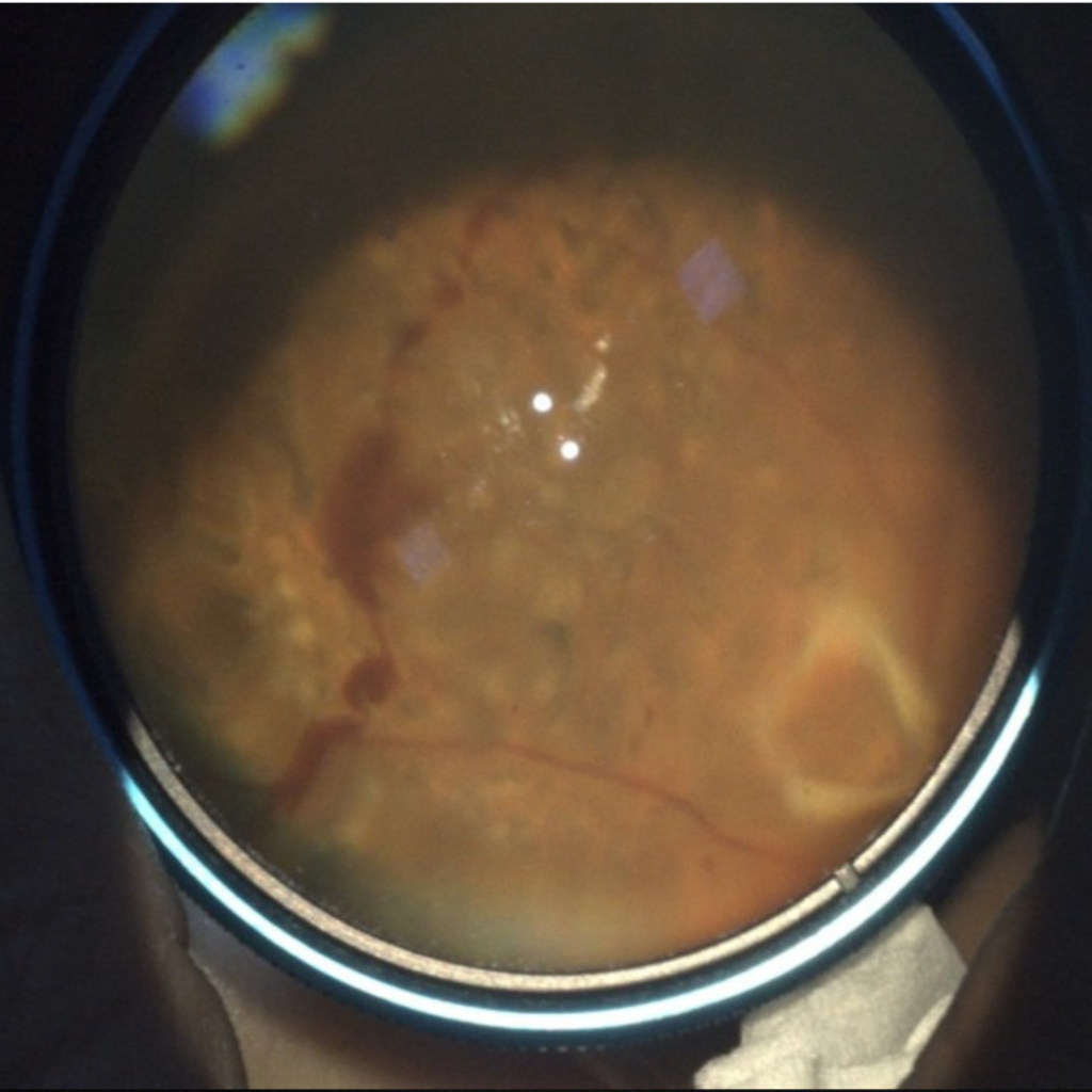
From theory to practice: proliferative diabetic retinopathy
Source: Dr Kenshuk Marwah
The potential value becomes easier to understand when we see the technology used in a real clinical case. In the Keeler Education Hub, we explore a case involving a patient with proliferative diabetic retinopathy and demonstrated the use of Vantage Plus Digital during fluorescein angiography.
The case helps demonstrate how a dynamic indirect examination and digital documentation can contribute another perspective when visualising retinal findings, particularly when there is clinical interest in areas beyond the posterior pole.
It also reinforces an important principle in diabetic eye care:
The value is not simply in capturing an image. It is in how the information contributes to understanding, documenting and managing the patient’s disease.
Finally, discover a range of images including Proliferative Diabetic Retinopathy and more in our: Image gallery
Diabetes Awareness Month reminds us that reducing avoidable vision loss is not about one examination, one image or one piece of equipment. It is about connecting the different parts of the patient’s journey.
At Keeler, we are proud to support eye care professionals with the tools, technology and education that help them examine, document and communicate the bigger picture to their collegues and patients.