


UBM Plus Connect – ultrasound probe and interface unit
Capture crystal clear images with a wide scope of imaging
Having trust and familiarity in your tools is crucial to performing ultrasound examinations with confidence. That’s why our ultrasound biomicroscipy (UBM) probe works on the same modality and uses similar measuring callipers as our B-scan probe, making it a valuage addition to your range of imaging equipment.
Designed for capturing high resolution images and videos of the anterior segment of the eye (including cornea, iris, ciliary body, zonules, crystaline lense and intraocular lens), the UBM probe is an easy-to-use portable device, ideal for both sharing between multiple users in a busy clinic, or using while on the go.
Working in harmony with Keeler Connect software (which can be plugged into any Windows™ laptop or desktop computer), UBM Plus Connect allows for easy set up and use straight away, meaning you can get started with examinations instantly.
The UBM probe is also fully compatible with the Keeler 4Sight™ ultrasound system – our all-in-one ultrasound solution.
Benefits:
- Unique focal zone guide for accurate results
- Safety guarded tip for peace of mind
- High quality imaging for reliable diagnosis
- Detailed structure definition – 48MHz scanning with a 60deg sweep angle
- Device portability – plugs into any Windows™ laptop or desktop computer
- All-in-one design to eliminate signal loss for sharper imagery
- Great value
- Competitive warranty and great customer support
- Fully compatible with Keeler 4Sight™ – giving you unmatched storage space
Accessories Included: Software installation, USB stick, probe holder, footswitch, wireless mouse, ophthalmic gel, and scleral shell.
![]()
This product is suitable for use by veterinarians. View our full range of veterinary instruments.
Description
The Keeler UBM probe makes capturing high resolution images and videos of the anterior eye simple!
Unique focal zone guide
Featuring a focal zone guide, the UBM probe helps you find the proper scanning technique during examinations, offering you accurate results every time so you can treat your patients with confidence.
Safety guarde tip
In order to provide peace of mind, it also comes with a guarded tip to prevent damage to the probe and discomfort to your patients.
Device portability
The UBM probe can be plugged into any Windows™ laptop or computer, making it perfect for sharing between multiple users or moving between exam rooms.
High quality imaging
Working at 48 MHz, the UBM probe takes high resolution images and videos over a 30º sweep angle, providing you with a wide scope of imaging and a detailed structure definition of the anterior segment of the eye.
Great value
At Keeler, we believe that value is more than the price alone. We look at every detail and quality components, results and high build quality shouldn’t cost the earth.
True partnership
True partnerships don’t stop, our industry-leading warranty and service programs, as well as our live help facility offer dependable support for the life of the product.
Features – Keeler Connect software:
-
- Intuitive interface
- Industry leading portability
- Ultrasound compatibility
- Great value
- Competitive warranty and great customer support
Features – Keeler UBM probe:
-
-
- High-Definition Imaging – Detailed structure definition including cornea, iris, ciliary body, zonules, crystalline lens, and intraocular lens, as well as pathologies
- State-of-the-art probe design – Sharper, more focused images due to the elimination of signal loss
- Easy to set up defaults – Smooth customer interface and automatic EMR exports that decrease examination time, increase patient throughput, and increase profit
- Unsurpassed data analysis – Contains tools for measuring sulcus-to-sulcus, anterior chamber depth, positioning of intraocular lenses, and filtration angle of the eye
- Adjustable video loop
- Fully upgradeable software
- EMR compatible
- DICOM ready
- Multiple measuring velocities for ACD/VCD
- PDF reporting
- Data Archive – Network or external location.
-
Specification:
-
- Weight: 6oz
- Dimensions: 7” long X 1.25” diameter
- Frequency: 48 MHz
- Axial Resolution: 0.015mm
- Electronic lateral resolution: 0.085mm
- Electronic gain: 0-112 dB
- Adjustable gamma: Linear, S-Curve, Log, Color
- Scanning angle: 30°
- Field of view: 32mm
- Frame rate per second: 10
- Sampling rate: 2048 (points per line)
- Vectors per frame: 256
- Focal point: 13mm
- Focal zone: 4mm
- TGC: Yes
- Frozen image gain adjustment: Yes
- Zoom: 2 x optical zoo
- Reports: PDF reporting
- Snapshot format: pdf, jpeg, bmp, png, tiff, gif
- Data archive/ Export capability: Yes
- Max. number of frames per scan: 256
- Size of cine loop: 13-128 MB
- Measurement calipers with velocity adjustment: 6 line, 2 area, 2 angle, 2 arrow
For all documentation associated with this product including cleaning instructions, product support and instruction manuals, please visit our support page here
Related products
-
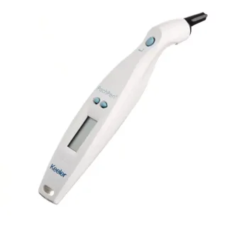
PachPen handheld pachymeter
24-5100/24-5100-V -
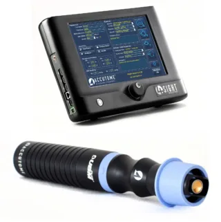
UBM ultrasound probe - 4Sight platform compatible
24-8000U / 24-8000U-V -
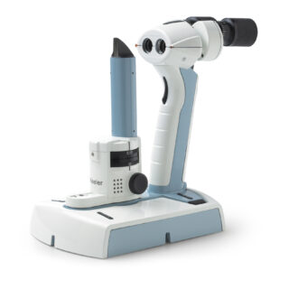
Keeler PSL Classic - Handheld portable slit lamp
3010-P-2000