


Simple Guide to the Most Common Optometry Equipment Names and Uses
If you’re considering optometry school or are an optometry school resident; understanding all of the most common optometry equipment can be slightly confusing and overwhelming. However, the experts at Keeler have created an intuitive and helpful resource tool to guide you along the way.
Direct Ophthalmoscopes
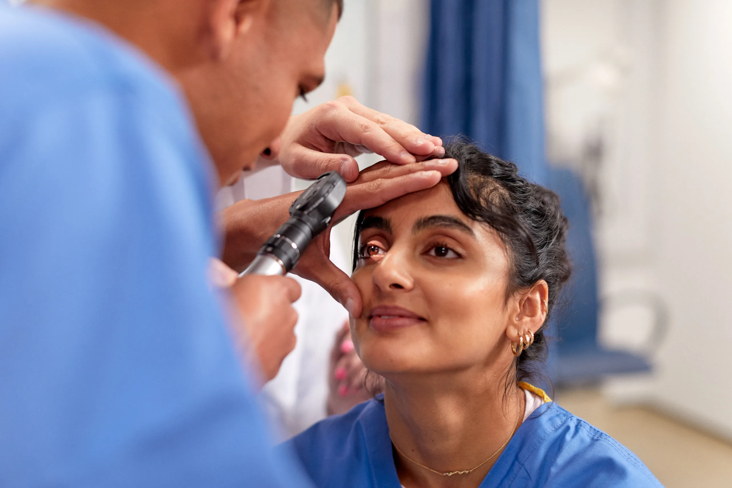
A direct ophthalmoscope is a handheld device used to look at the inside of the patient’s eye. Direct ophthalmoscopes utilize a set of lenses that focus on certain eye structures and use batteries to create light.
With direct ophthalmoscopy, the physician looks through one of the small lenses into the eye to view the appearance of the:
- arrow-rightCornea,
- arrow-rightLenses,
- arrow-rightVitreous humour,
- arrow-rightAqueous humour, and
- arrow-rightSurface of the retina.
Direct ophthalmoscopes allow practitioners to see intricate details with the ability to utilize magnification of around 15x.
Binocular Indirect Ophthalmoscope
The binocular indirect ophthalmoscope is a headset device the practitioner wears. This device is used with a condensing aspheric lense placed close to the patient’s eye. The Binocular Indirect (BIO) allow practitioners to observe the entire retina and have access to wider field of view in comparison to the direct ophthalmoscope.
As a result, BIOs usually have magnification of around 5x. Some BIOs — like the Vantage Plus Digital System — are used for training and include a built-in video camera, so students can effectively observe. In August 2017 The National Board of Examiners in Optometry exclusively uses the Keeler Vantage Plus Digital System for part III clinical skills exams. You can also find a wide range of additional BIO accessories for multiple uses.
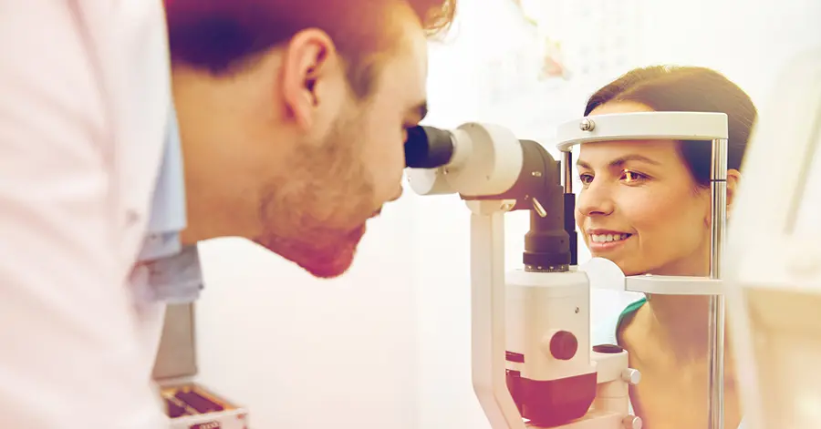
Slit Lamps
Slit lamps are table-mounted instruments or hand-held slit lamps used to look into a patient’s eyes with bright light and a microscope. Slit lamps can have a range of magnification strengths from 6x up to 40x. The beam of light can be modified in a variety of ways, including:
- arrow-rightHeight
- arrow-rightIntensity
- arrow-rightAngle
- arrow-rightWidth
- arrow-rightColor
- arrow-rightDirection
Patients can gently rest their chin and forehead against it slit lamp device, and the eye doctor can examine their eyes from the other side. Different types of slit lamps can detect deeper and more serious issues such as cataracts, corneal lesions, or corneal ulcers.
Surgical Loupes
Surgical loupes are telescopic additions to glasses or goggles commonly used by physicians and practitioners. Surgeons especially are seen with surgical loupes during surgery, but any doctor can use them to take a closer look for any reason, including inspecting a patient’s eyes.
For eye doctors and eye surgeons, surgical loupes are quite useful in examining the surfaces of parts of the eye, including the surface of the cornea. Eye surgeons also typically wear surgical loupes during certain types of eye surgery.
Retinoscopes
A retinoscope is a hand-held device used for looking at a patient’s retinas and measuring errors in the reflection. Retinoscopy is a useful technique for prescribing corrective lenses; typically the test produces a good baseline for narrowing down the exact prescription via a subjective refraction test.
Pachymeters
A pachymeter is a device for measuring the thickness of the corneas of a patient’s eyes. These devices are offered in a variety of shapes and sizes. While the handheld pachymeter is quite common, you’ll also find a wide range of tabletop instruments. Pachymeters are useful when a practitioner suspects a patient has glaucoma as well as an important measurement tool to use prior to eye surgery.
Tonometers
A tonometer is an advanced medical device for measuring ocular pressure. Similar to pachmeters, there are a vast range of different types of tonometers available in different shapes and sizes for various techniques and tests:
- arrow-rightStandard contact tonometers and digital contact tonometers that touch the eye,
- arrow-rightNon contact tonometers that do not touch the eye at all,
- arrow-rightTable-mounted tonometers, and
- arrow-rightHand-held contact tonometers.
In either case, the tonometer is used to perform tonometry that tests the fluid pressure inside a patient’s eyes. It’s another useful screen when a patient is suspected of developing or at risk for glaucoma.
Ophthalmic Cryosystems
Cryotherapy devices are used in ophthalmology to treat a range of conditions and disorders of the eye. Ocular cryotherapy explains the extremely frigid temperatures used to treat conditions of the eyelids or eyes. Innovative cryotherapy techniques have been used since the mid 1960s. These treatments are typically utilized as a surface technique.
Some of the common applications of ophthalmic cryosurgery include:
- arrow-rightCryoextraction of cataracts
- arrow-rightCyclocryopexy for advanced glaucoma
- arrow-rightRetinal cryopexy
- arrow-rightFreezing of lash roots for recurrent trichiasis
- arrow-rightPeripheral retinal cryoablation for neovascular glaucoma
- arrow-rightCryosurgery for conjunctival neoplasias of the epithelium
- arrow-rightCryotherapy for malignancies of the eyelids
- arrow-rightPeripheral retinal cryoablation
- arrow-rightCryoablation of retinoblastomas
Contact Keeler
If you’re looking for the newest and most reliable ophthalmic instruments, look no further than Keeler. For more than 100 years, Keeler has been manufacturing equipment and providing the industry with thought leadership.
Contact Keeler to learn more about any type of the most common ophthalmic equipment.