


Bringing 5 of the Most Common Eye Diseases into Focus
Unfortunately, many of the most common eye diseases have no early warning signs, are painless, and results in no vision change until the disease has significantly advanced. However, optometrists, ophthalmologists, and vision practitioners are on the front line of defense to protect patients against the following most common eye diseases.
Glaucoma
As the second leading cause of blindness, glaucoma has caused six million people in the world to suffer blindness in both eyes from glaucoma. Based on one estimate, over three million people in the United States suffer from glaucoma.
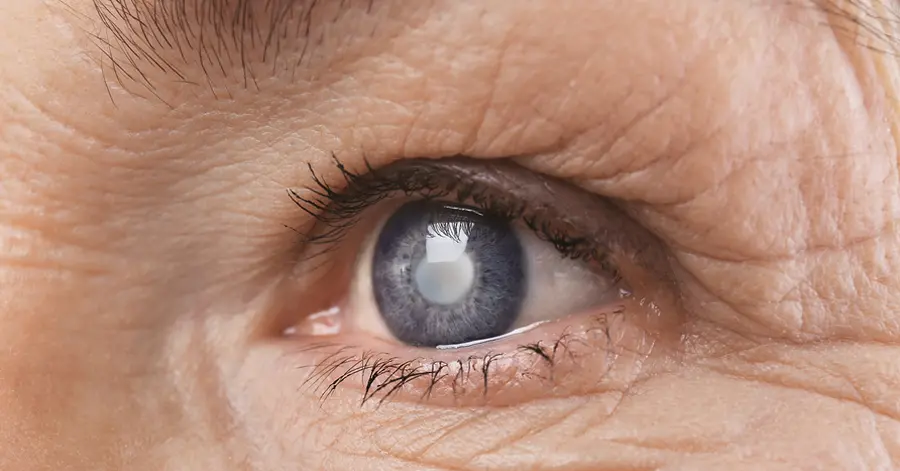
Glaucoma causes damage to the optic nerve of the eye. This degenerative disease continues to worsen over time. Because glaucoma is associated with a buildup of eye pressure, it usually doesn’t show up in patients until later in life and tends to be inherited.
Overtime, this increased pressure — also called intraocular pressure — results in optic nerve damage. The optic nerve plays a vital role of transmitting images to the brain. If the high pressure and damage to the optic nerve goes unchecked, the glaucoma will lead to the patient suffering permanent vision loss. As a result, it’s imperative for ophthalmologists to utilize tonometry devices to test the inner eye pressure, such as:
Cataracts
Characterized by a white, cloudy lense in the eye, cataracts are more common in those over the age of 50. While cataracts are definitely age related, they can develop in patients at any age. Cataracts can be the result of UV exposure, injury, or protein deterioration over a period of time. If cataracts are not treated in a timely manner, it can result in severe vision loss.
Fortunately, cataracts are a very common problem that can be rectified through surgery with ophthalmic instruments like the A-Scan Plus Connect. Whether surgery is required depends on how much it affects the patient’s quality of life and the degree of vision loss.
To test for cataracts, the patient’s eyes must be dilated, which makes it easier for you to see the back of their eye. Once dilated, the doctor uses either a slit lamp or an ophthalmoscope to inspect signs of cataracts.
Diabetic Retinopathy
Patients who have prolonged high blood sugar from Type 1 or Type 2 diabetes can suffer from diabetic retinopathy. If left untreated, diabetic retinopathy can cause blindness. This common eye disease is caused when too much blood sugar changes the blood vessels in the back of the eye. This prevents the retina from getting an adequate amount of nutrients required to properly maintain vision. Any of your patients who have Type 1 or Type 2 diabetes is at risk to develop diabetic retinopathy. Factors that affect the patient’s risk include:
Significant vision loss from diabetic retinopathy can usually be prevented with laser surgery. Laser photocoagulation can destroy or seal any leaking or growing retinal blood vessels.
Color Blindness
Virtually every person who suffers from color blindness can see colors, but they have problems distinguishing between certain colors. Everyone who has color blindness doesn’t have problems with the same color.
Some people cannot determine yellows from blues; most cannot distinguish between greens and reds; and a small group can only see white and black, which is a condition called monochromatism. Determining whether a patient has color blindness involves a series of different sight and color vision testing.
Keratoconus
Typically, the clear outer lens of the eye — the cornea — has a ball, dome-like shape. In some patients, however, the collagen holding the cornea in place gets weak and causes it to become cone shaped, which is a condition called Keratoconus.
Keratoconus can result in significant vision loss if it’s not treated quickly and early. Optometrists and ophthalmologists utilize a pachymeter to determine the thickness of the patient’s cornea. This device is utilized to conduct corneal pachymetry before a refractive surgical procedure or Keratoconus screening.
Treating Keratoconus usually starts with eyeglasses followed by prescribing gas-permeable, rigid contact lenses to improve vision and strengthen the cornea. Another option to help prevent the progression of Keratoconus is cornea collagen crosslinking. Implants placed underneath the cornea surface or intacs may be used to improve vision and reduce the cone shape.
Contact Keeler Ophthalmic Instruments to Treat the Most Common Eye Diseases
For more than 100 years, Keeler Ophthalmic Instruments has been on the precipice of innovation. With an unwavering focus on the future, Keeler invests heavily in research and development to advance the field. We pride ourselves on purposeful innovation centered on initiatives of customer involvement. We make sure to include customers throughout the design process to guarantee our equipment meets your needs and exceeds your expectations.
Contact Keeler Ophthalmic Instruments today to learn more about any of our world-class equipment used to treat the most common eye diseases.