


Seeing beyond the clouded lens with ocular ultrasound
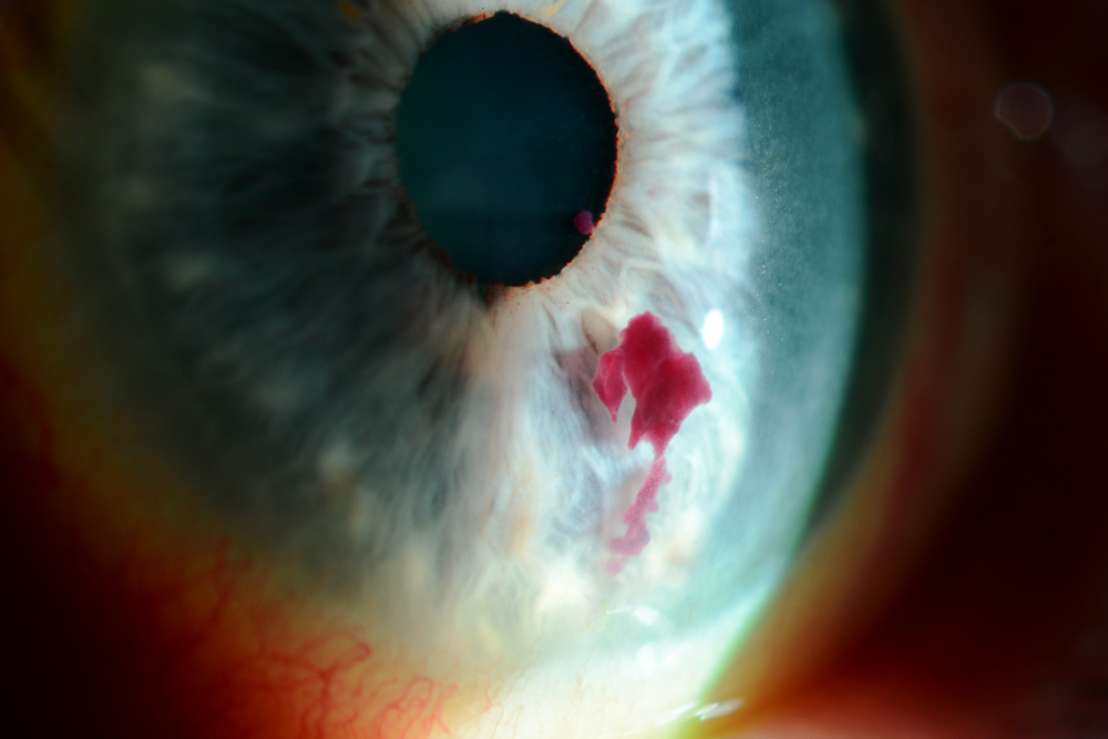
Cataracts are a leading cause of visual impairment worldwide, clouding the lens of the eye and obstructing clear vision. While cataract surgery is a common and highly effective treatment, assessing the underlying structures of the eye becomes challenging when the lens is opacified. This is where ocular ultrasound, a non-invasive imaging technique, steps in as a crucial diagnostic tool.
For Cataracts Awareness Month, we’ll review how ocular ultrasound helps ophthalmologists identify cataracts and help prevent blindness.
What is ocular ultrasound?
Ocular ultrasound, also known as B-scan ultrasonography, uses high-frequency sound waves to create detailed images of the eye’s internal structures. It is especially valuable in cases where direct visualisation is hindered, such as with dense cataracts. By providing a clear view of the posterior segment of the eye, ocular ultrasound helps clinicians both in ophthalmology and optometry evaluate and manage various eye conditions effectively.
Why use ocular ultrasound for cataracts?
When cataracts block the view, it is essential to ensure that there are no other underlying issues that could affect the surgical outcome of a patient’s vision post-surgery.

Assessing the retina and vitreous:
Cataracts can obscure the retina and vitreous humor, making it difficult to detect abnormalities such as retinal detachments, vitreous haemorrhages, or tumours. Ocular ultrasound penetrates through the opaque lens, providing clear images of these structures.
Evaluating optic nerve health:
The optic nerve head is critical for vision. Ultrasound helps in assessing optic nerve anomalies, ensuring that any optic neuropathy is identified before cataract surgery.
Detecting posterior segment disorders:
Conditions like posterior vitreous detachment, choroidal detachments, and intraocular foreign bodies are detectable via ocular ultrasound, aiding in comprehensive preoperative assessment.
Advantages of ocular ultrasound
- Non-invasive:
The procedure is safe and painless, posing no risk of radiation exposure. - Real-time imaging:
Provides immediate results, allowing for quick diagnosis and decision-making. - Detailed visualisation:
Offers clear images even when the view is obstructed by cataracts, facilitating thorough assessment.
Understanding A-scan and B-scan ultrasound
In ocular ultrasonography, both A-scan and B-scan techniques can be used to obtain comprehensive information about the eye’s condition.
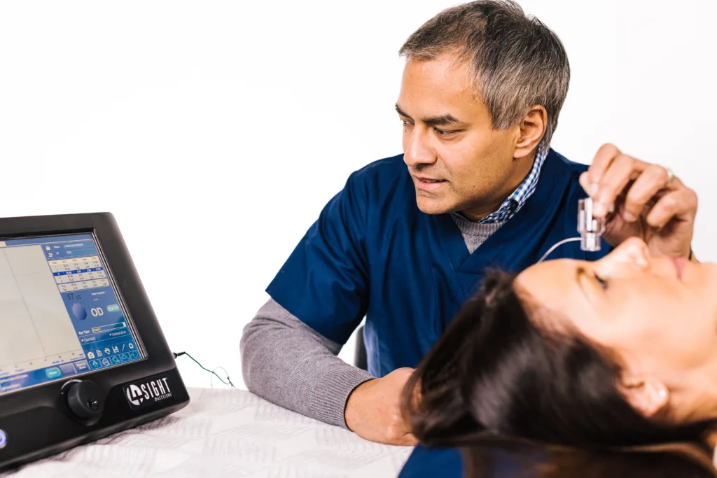
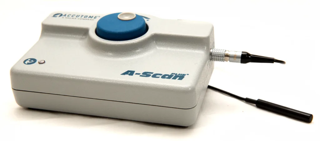
A-scan ultrasound:
A-scan (amplitude scan) ultrasound measures the eye’s axial length and provides a one-dimensional waveform that represents the distances between internal eye structures. This information is crucial for calculating the appropriate intraocular lens (IOL) power for cataract surgery and also helps with clinical assessment and diagnosis.
B-scan ultrasound:
B-scan (brightness scan) ultrasound creates two-dimensional cross-sectional images of the eye, allowing detailed visualisation of the retina, vitreous, and optic nerve head. This scan is essential when the view is obstructed by cataracts or other opacities.

Conclusion
Ocular ultrasound is an indispensable tool in modern ophthalmology and optometry for many reasons, but particularly in managing patients with cataracts. By providing a window into the eye’s interior when direct visualisation is not possible, it ensures that comprehensive and informed care is delivered.
For patients awaiting cataract surgery, ocular ultrasound offers peace of mind, knowing that their eyes have been thoroughly evaluated, allowing for a successful outcome with the best possible vision after surgery.
In the ever-evolving technical field of eye care, ocular ultrasound is an example of how technology can overcome visual barriers, ensuring that no detail goes unnoticed.
Keeler offers a wide range of ultrasound products designed to support eye health and help prevent vision loss, all around the world. With their user-friendly, ergonomic design, Keeler’s ultrasound devices enable clinicians in both ophthalmology and optometry to provide the best possible care.