


How UBM imaging is key for eye care professionals
Ultrasound Biomicroscopy (UBM) imaging is a technique for ocular pathology diagnosis. By producing a very high frequency, UBM imaging delivers greater quality imaging at extremely close distances — allowing for micro-sized images for specific diagnoses.
Due to its high resolution, UBM imaging doesn’t typically penetrate as deeply as other types of ultrasound techniques. But for the anterior portion of the eye, UBM is extremely effective.
With the right UBM imaging equipment, experienced examiners can make quicker and more accurate diagnoses, with greater comfort and convenience for the patients.
At Keeler, you can browse a full range of high-quality, durable UBM imaging devices, including:
What is UBM imaging?
Ultrasound Biomicroscopy (UBM) imaging provides an in depth cross-section of the eye at a microscopic level. UBM imaging uses drastically higher sound wave frequency than either A-scan technology or B-scan equipment.
While the image resolutions are much greater, the ability of UBM imaging technology to penetrate tissue is diminished. As a result, UBM imaging is typically utilized to produce images in the anterior section of the eye as well as help with diagnosing a vast range of eye diseases.
What does UBM imaging do?
UBM imaging has the inherent advantage of detecting anterior ocular segment structures quite clearly, and without any invasive discomforts. Structures visualized include:
In addition, UBM imaging is regularly used to diagnose a vast range of eye diseases, including (but not limited to) glaucoma, ciliary body cysts, and angle trauma.
Applications of ultrasound biomicroscopic imaging
UBM imaging can be an effective alternative to gonioscopy, which is used to evaluate the anterior eye angle and drainage system. Gonioscopy can be uncomfortable to patients and is almost entirely subjective to the examiner.
UBM imaging allows for more objective, quantitative measurements. In clinical research, UBM is often employed to learn more about the angle and to find more quantitative analysis of the anterior chamber angle.
Standard clinical UBM imaging measurements can include:
These measurements are often used to monitor and track changes in the anterior chamber angle over time as well as evaluate post-operation success in glaucoma patients.
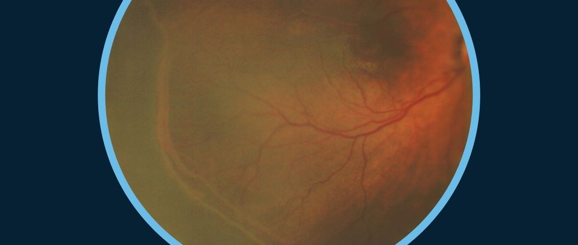
UBM imaging of the cornea
With UBM imaging of the cornea, pathologies are more easily and more accurately diagnosed, including:
Procedural advantages of UBM
One of the most popular aspects of ultrasound biomicroscopic imaging is its non-invasive nature. Ultrasonic waves naturally avoid pigmentation, opaque eye structures, and other barriers to light-based imaging, giving UBM a clear advantage in this regard.
With the immersion shell method, which is very much like the method for A-scan imaging, test time is short and an anesthetic eye drop helps patients avoid any discomfort. On the other hand, the Clear Scan method allows patients to sit up without the use of anesthetic eye drops.
Anterior segment imaging with UBM provides essential data to ophthalmologists, as well as the visual aid to evaluate and assess patients’ eyes for for abnormalities like tumors, cysts and trauma. This data is not only key in detection and diagnosing, it is also effective in follow-up treatment and monitoring over time.
Ocular abnormalities are easily detected with UBM imaging, and early detection is available thanks to this technology, assisting in treatment success by alerting doctors and patients and inspiring preventative care.
Contact Keeler for UBM Imaging
For more than 100 years, Keeler has led the way in ophthalmic imaging. We offer multiple UBM imaging devices and proudly stand behind all of our equipment with industry-leading warranties and extended service.