Reliability at high-altitude: An oculomics study up Mt Kilimanjaro
Keeler
Reaching new heights: Why Keeler equipment joined a Mount Kilimanjaro expedition to help advance the future of medical research.
The human eye may reveal far more about our health than we once imagined.
In this blog, we’re thrilled to share the story of an expedition that set out to explore how high-altitude environments affect the human body, with a particular focus on the retina as a window into systemic health.
Led by Ophthalmologist Dr. Jennifer Grin, the study brought together cutting-edge research, portable medical technology, and the challenging conditions of one of the world’s most iconic mountains.
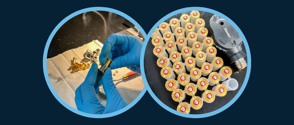
To support the research team, Keeler provided equipment, including a Portable Slit Lamp, enabling clinicians to perform detailed ocular examinations even in a remote, high-altitude environment.
Throughout the expedition, researchers collected retinal images and physiological data from climbers as they ascended the mountain. The results offer valuable insights into how the body adapts to reduced oxygen levels and highlight the potential of oculomics in detecting systemic disease.
Read on as Dr. Jennifer Grin shares insights from the expedition, and discusses how our portable ophthalmic equipment played a critical role in making this research possible.
Q&A with Dr. Jennifer Grin: Exploring Oculomics at high altitude
“The retina contains an extraordinary amount of information about our health, and now with modern imaging and AI, we’re only beginning to understand all that it can reveal” – Dr. Jennifer Grin
1: What is oculomics, and why is it significant?
Oculomics is an emerging field that combines advances in ocular imaging, big data, and artificial intelligence to detect systemic diseases through the eye.
Recent technological developments, including portable imaging devices and AI-driven analysis allow researchers to identify subtle patterns in retinal images that may signal diseases such as stroke, cardiovascular disease, and Alzheimer’s, often before clinical symptoms appear.
This approach has the potential to transform how we detect and monitor disease, offering opportunities for earlier diagnosis and improved patient outcomes.
2. What was the overall purpose of the study being conducted at altitude?
The study aimed to better understand how the human body responds to hypoxia, or low oxygen levels, which occur naturally at high altitude. By capturing retinal images alongside physiological measurements, researchers were able to study how the body adapts as oxygen levels decrease during ascent.
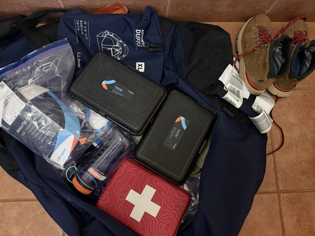
The expedition also demonstrated the feasibility and value of portable ophthalmic and medical equipment in remote expedition settings.
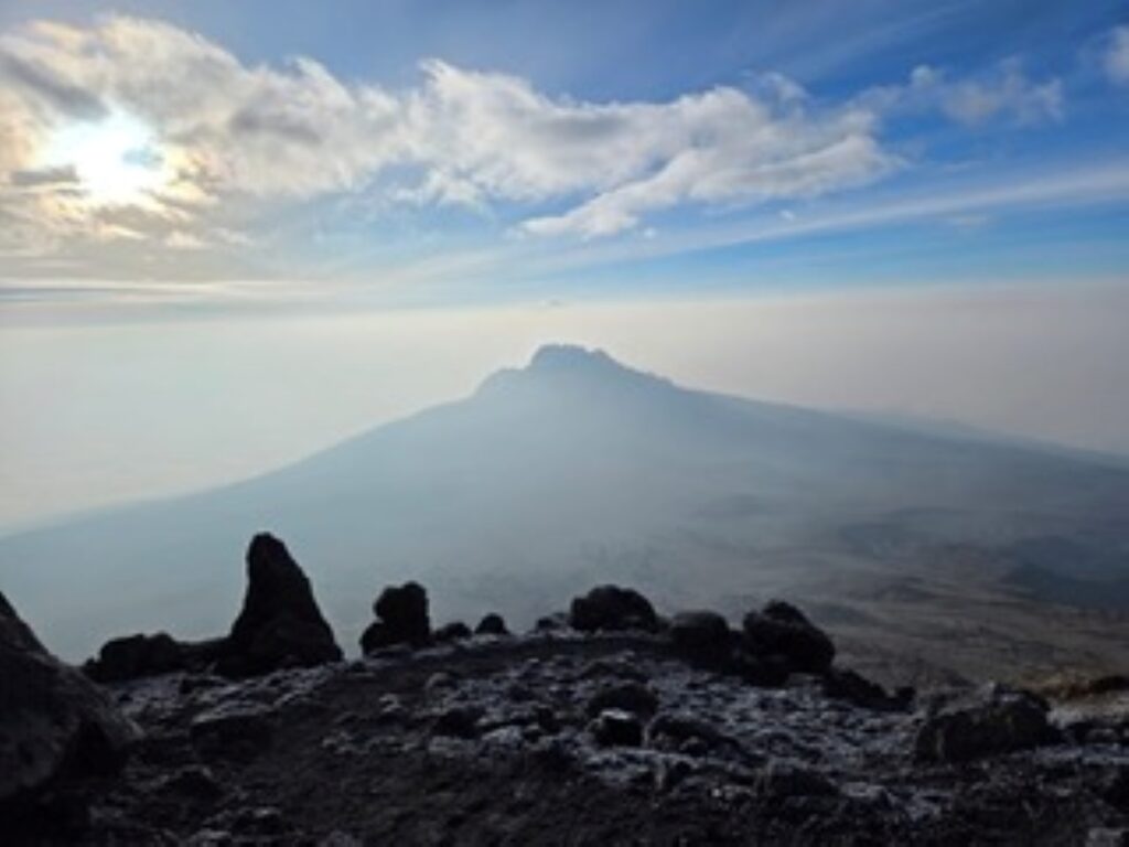
3: Why is high altitude considered a valuable environment for medical research?
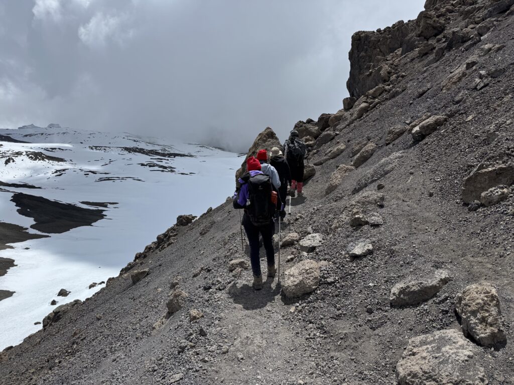
High altitude serves as a natural laboratory for studying hypoxia.
As elevation increases, oxygen levels decrease, creating conditions similar to those seen in many medical conditions and diseases.
Studying these effects in a real-world environment provides unique insight into how the body responds to oxygen deprivation and helps researchers better understand the physiology behind altitude sickness and other hypoxia-related conditions.
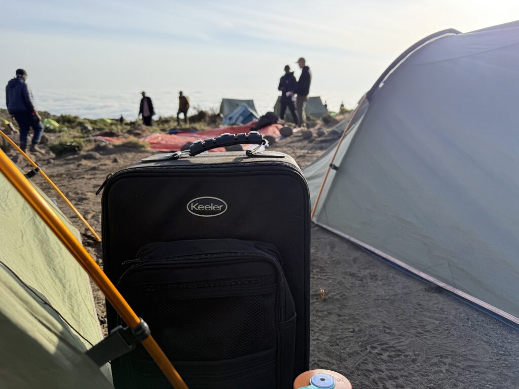
6. In practical terms, how was the equipment used during the expedition?
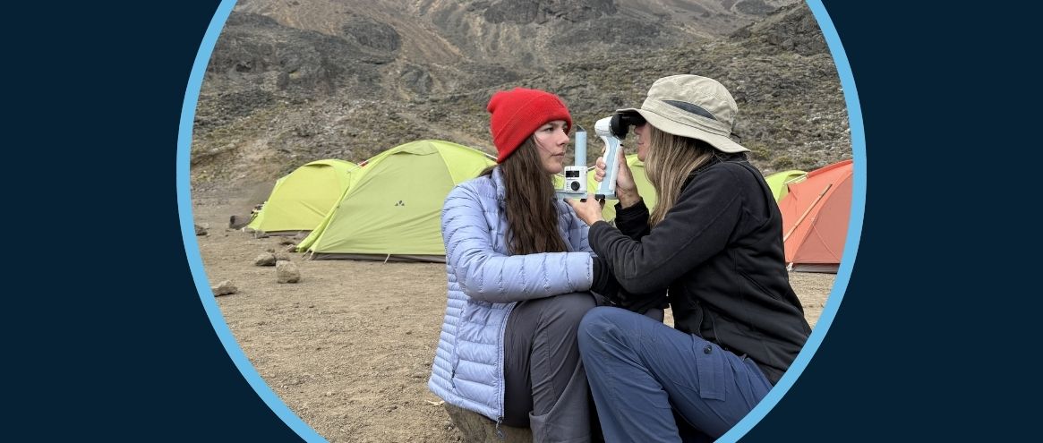
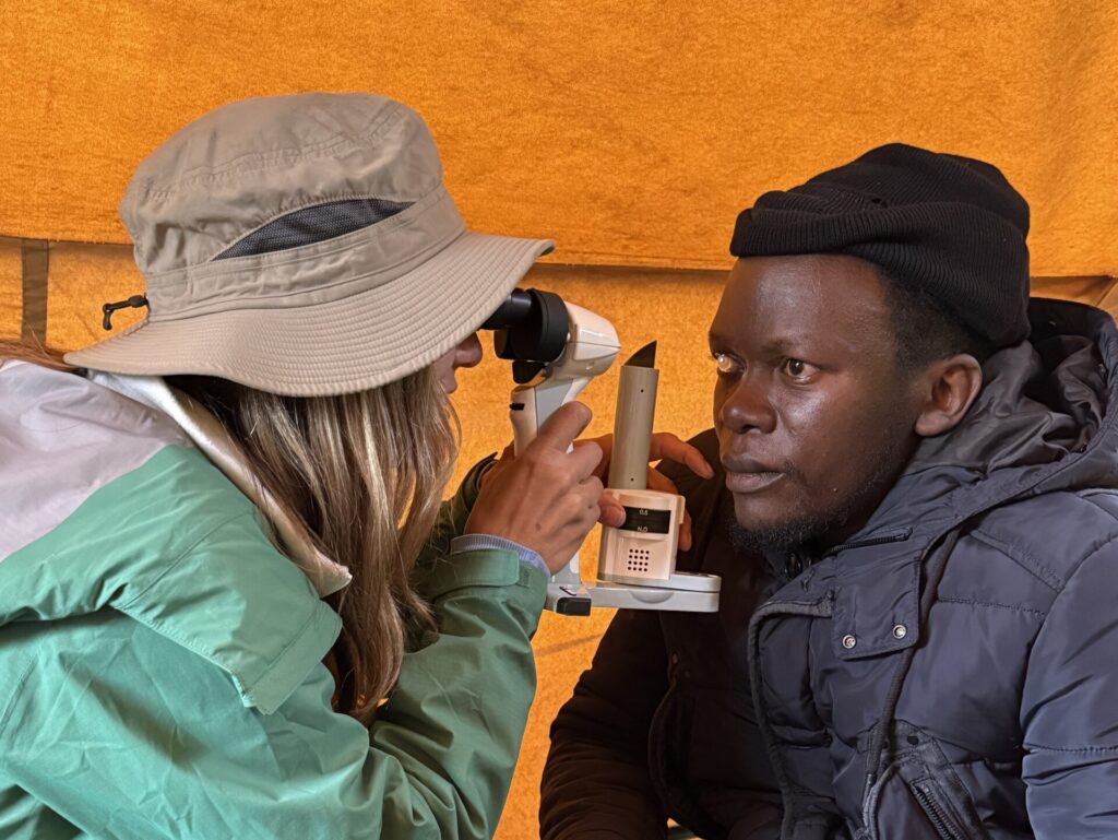
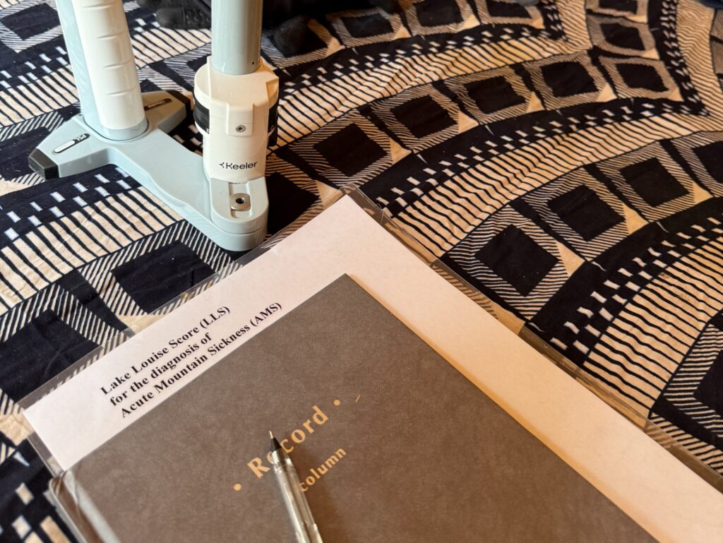
At the end of each day’s climb, researchers established a makeshift medical tent where participants were evaluated.
A wide range of portable medical devices were used to collect biometric data, including: Oxygen saturation, Heart Rate measurements, Electrocardiography (EKG), Lung and ocular ultrasound, EEG brain monitoring and Fundus photography.
Having reliable, portable diagnostic equipment allowed the team to deliver medical care and collect research data even in remote, high-altitude conditions.
4. What role does the eye play in studying systemic health and hypoxia?
The eye is the only place in the human body where blood vessels and nerves can be directly visualised non-invasively.
Because the retina is essentially an extension of the brain, it offers a unique window into both vascular and neurological health.
Changes in retinal blood vessels can provide valuable clues about how the body responds to stressors such as hypoxia.
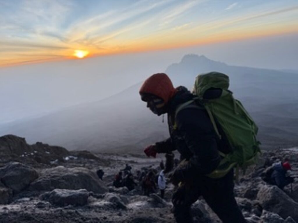
5. Who were the study participants?
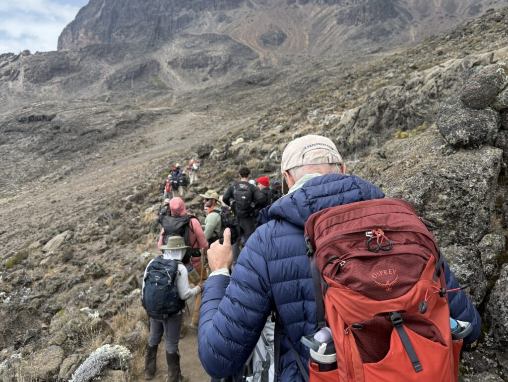
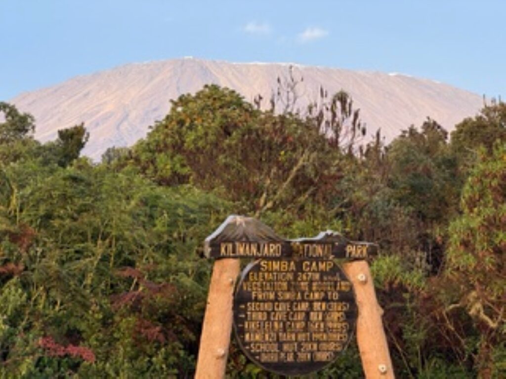
Twenty climbers between the ages of 16 and 68 participated in the study during the seven-day expedition on Mount Kilimanjaro in Tanzania.
Most participants were lowlanders travelling to high altitude, though local climbers and physicians also took part in the research.
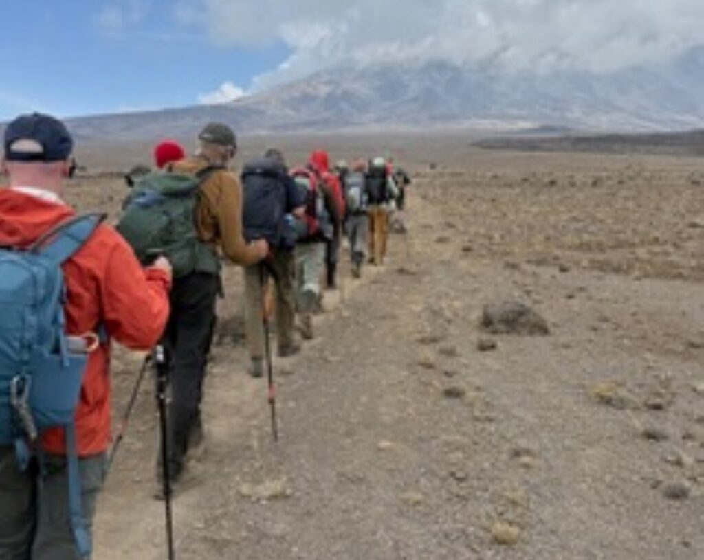
7. What process did you use for data collection during ascent?
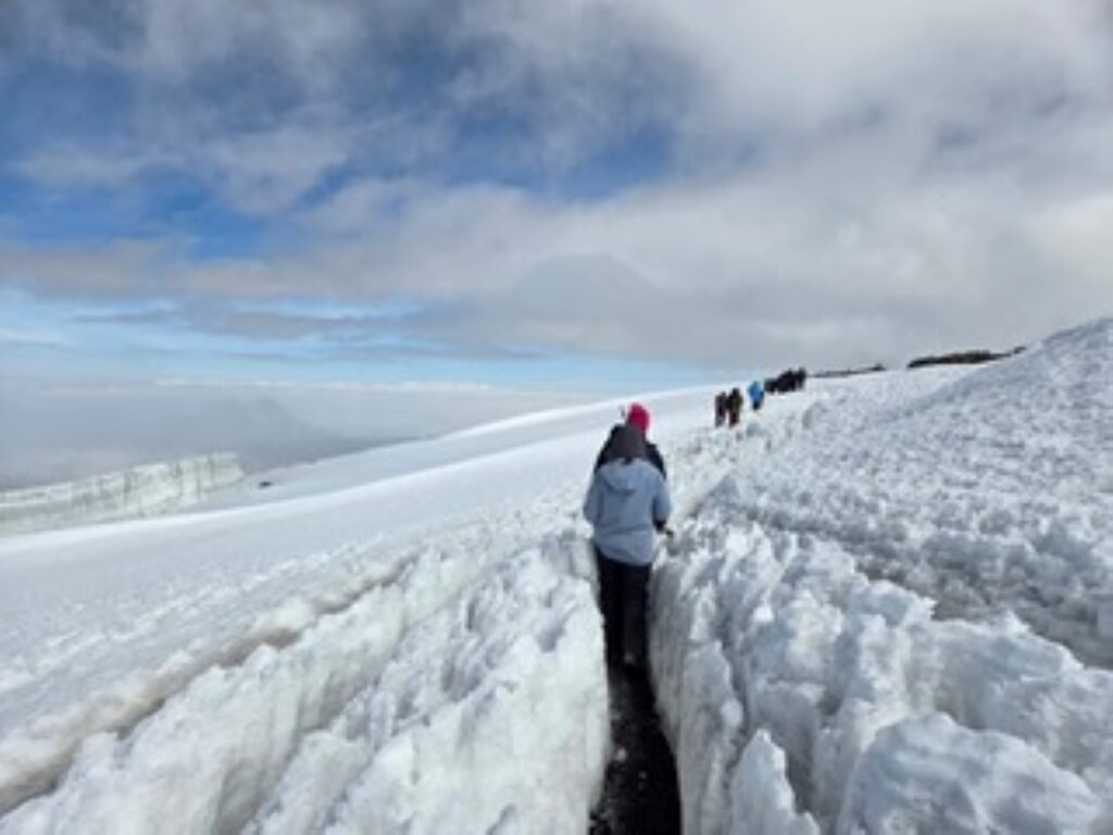
Measurements and retinal imaging were performed at Base Camp (approximately 1,500 metres) and at each ascending camp prior to the summit attempt.
This allowed researchers to document retinal vascular and optic nerve changes as altitude increased.
For safety reasons, no measurements were collected at the summit (19,300 feet / 5,882 metres).
8. How was artificial intelligence (AI) used in this research?
Artificial intelligence was used to automate the analysis of retinal images captured during the expedition.
AI algorithms extracted key retinal vessel metrics, including vessel diameter and vessel tortuosity, from images taken with a handheld fundus camera.
AI systems can also detect subtle retinal patterns invisible to the human eye.
For example, some models can differentiate between male and female retinas, something human ophthalmologists have not reliably been able to do.
This highlights the vast amount of information embedded within retinal images, which is providing incredible insights to the early detection of disease.
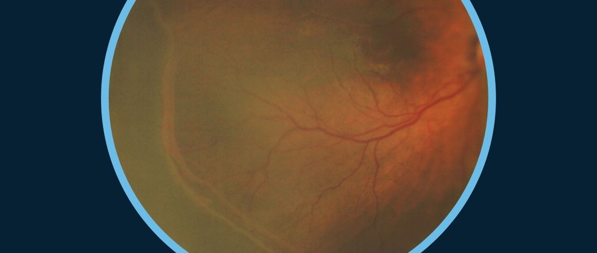
9. What physiological eye changes were observed at altitude?
Researchers observed dilation of retinal blood vessels and increased vessel tortuosity at higher altitudes.
These changes indicate increased blood flow as the body attempts to deliver more oxygen to the brain and eyes in hypoxic conditions.
In some participants, retinal hemorrhages were also observed as part of high altitude illness.
10. How did the equipment perform in high-altitude conditions?
Despite the challenging environment, the portable equipment performed exceptionally well.
Reliable diagnostic tools were essential for conducting research in such a remote location, and the equipment allowed clinicians to perform detailed examinations even under demanding conditions.
11. What role did PSL Classic play during the expedition?
It enabled clinicians to quickly assess symptomatic individuals for conditions such as eye irritation, infection, foreign bodies, redness, and inflammation, allowing for prompt diagnosis and treatment.
In a remote expedition setting, having access to a high-quality portable slit lamp proved invaluable.
12. Were altitude sickness conditions observed in the study?
Yes.
High altitude illness is common in individuals ascending above 2,500 metres and can range from mild to the potentially life-threatening conditions of High Altitude Pulmonary (HAPE) and High Altitude Cerebral Edema (HACE).
Participants were assessed daily by the medical team and a specialized questionnaire was used to document symptoms of headache, nausea, dizziness, and sleep disturbances.
One patient developed retinal hemorrhages as part of High Altitude Retinopathy but thankfully no one developed either HAPE or HACE.
Combining these clinical assessments with physiological and ocular measurements may help researchers better understand the underlying mechanisms of altitude illness.
13. Was there an educational component to the expedition?
Yes. The research team collaborated with Marangu Hospital in Tanzania and introduced portable ophthalmic equipment to local clinicians who previously had limited access to such tools.
This included hands-on demonstrations to support community outreach and continued medical education.
Dr. Goodluck Malle, Chief Medical Officer, Marangu hospital
“High on the mountain, the retina allows us to see what the body endures“ – Dr. Jennifer Grin
Credits & Thanks:
We’re incredibly proud that our ophthalmic devices accompanied the team on this ambitious ascent, contributing to research that will advance medical understanding for years to come.
Our sincere thanks to Dr. Grin for taking us behind the scenes of this high-altitude journey, and for her ongoing progress in this important research.
Jennifer Grin, MD, FACS , is a Yale University, board-certified ophthalmologist and Fellow of the American College of Surgeons, with more than 25 years of clinical and surgical experience. Based in the Denver metropolitan area, she built and led a thriving practice as a high-volume cataract surgeon, serving patients across the Rocky Mountain region.
With a passion for both medicine and exploration, her career blends clinical excellence with scientific discovery. Dr. Grin’s interest in high-altitude ophthalmology has taken her from Everest Base Camp to the summit of Mount Kilimanjaro. She is now focused on advancing Oculomics, a cutting-edge field at the intersection of eye and systemic health, thus redefining the eye as one of the most powerful diagnostic windows into human health and the future of precision medicine.
All photos used with the kind permission of Dr. Jennifer Grin.


")


")