As we near the end of Glaucoma Awareness month, we’d like to reflect on the fact that glaucoma is often called the “silent thief of sight”, because this condition can progress without symptoms until irreversible vision loss occurs.
Whilst routine eye examinations remain paramount, as they are the foundation of early detection and ongoing monitoring, modern technology allows us to go further, giving clinicians powerful tools to detect glaucoma earlier, manage it more effectively, and educate patients with confidence.
In this blog, we explore the imaging techniques used today for an effective diagnosis and care plan.
What is Glaucoma?
Glaucoma is a group of eye conditions that damage the optic nerve, often due to elevated intraocular pressure (IOP).
It can affect anyone, usually after adulthood, and is frequently symptomless in its early stages, making awareness and early detection crucial.
Types of Glaucoma
Glaucoma isn’t a one-size-fits-all condition. It presents in several forms, each with unique characteristics and management needs:
Primary Open-Angle Glaucoma (POAG) – The most common type, often symptomless until advanced stages, making regular eye exams critical.
Angle-Closure Glaucoma – A sudden, severe form that can cause pain and vision loss quickly, requiring urgent intervention.
Normal-Tension Glaucoma – Optic nerve damage occurs even when IOP is within normal limits, highlighting the importance of comprehensive imaging and monitoring.
Secondary Glaucoma – Linked to other eye conditions, trauma, or systemic disease, adding complexity to diagnosis and treatment.
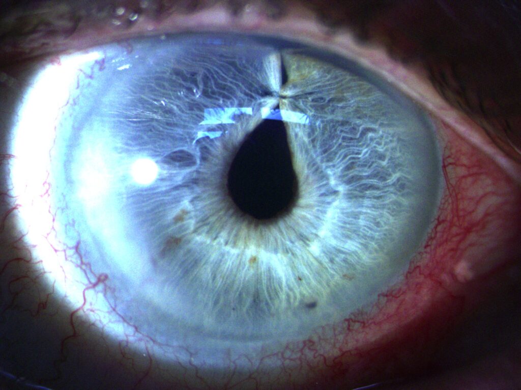
Iridoplasty
For eye care professionals, understanding these variations is key to both leveraging advanced imaging for an accurate diagnosis and tailored care.
Why imaging matters in Glaucoma care
Although traditional tests like IOP measurement and visual fields remain essential, they don’t always reveal early structural changes. And that’s where imaging steps in:
Anterior segment imaging has become a cornerstone of advanced glaucoma care. Here are the benefits of the options available:
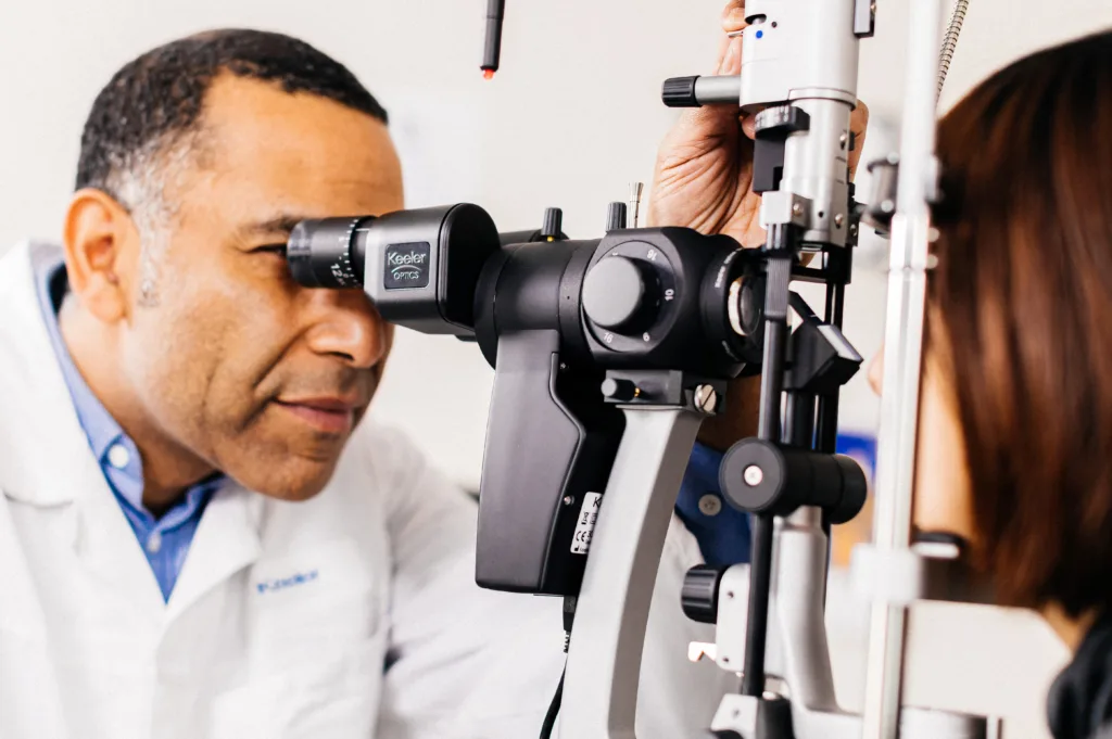
Digital slit lamp imaging provides crystal-clear views of the anterior chamber and optic nerve head, enabling documentation and monitoring.
Van Herick’s assessment and Gonioscopy remain vital for angle evaluation, but imaging adds precision and provides a permanent record for comparison over time.
Ultrasound Biomicroscopy (UBM) goes deeper, offering cross-sectional views beyond the iris to reveal structures like the ciliary body and zonules. This is invaluable for diagnosing narrow angles, plateau iris, and secondary glaucoma’s.
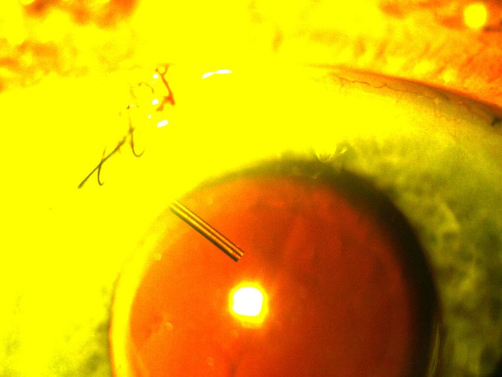
XEN migration in AC - shiny yellow
2. OCT and Fundus photography for routine monitoring
In addition to anterior imaging for angle assessment, OCT and fundus photography remain essential for monitoring optic nerve health and retinal nerve fibre layer changes.
These technologies complement IOP, Visual Fields, slit lamp and UBM imaging, ensuring comprehensive glaucoma management.
Glaucoma eye plate + RS
3. Slit lamp imaging in surgical care
For procedures like Minimally Invasive Glaucoma Surgery (MIGS), slit lamp imaging supports both pre-operative planning and post-operative monitoring.
High-resolution images confirm implant positioning and help educate patients with clear visuals that build trust and compliance.
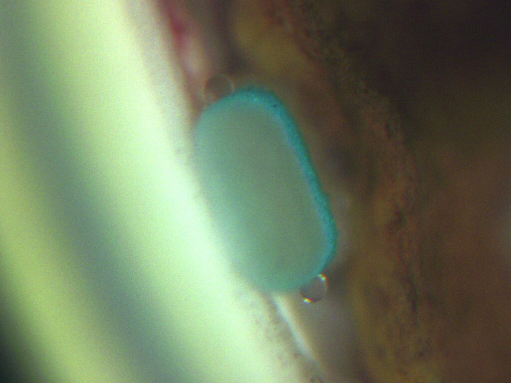
Rare MINIject with 2x droplet
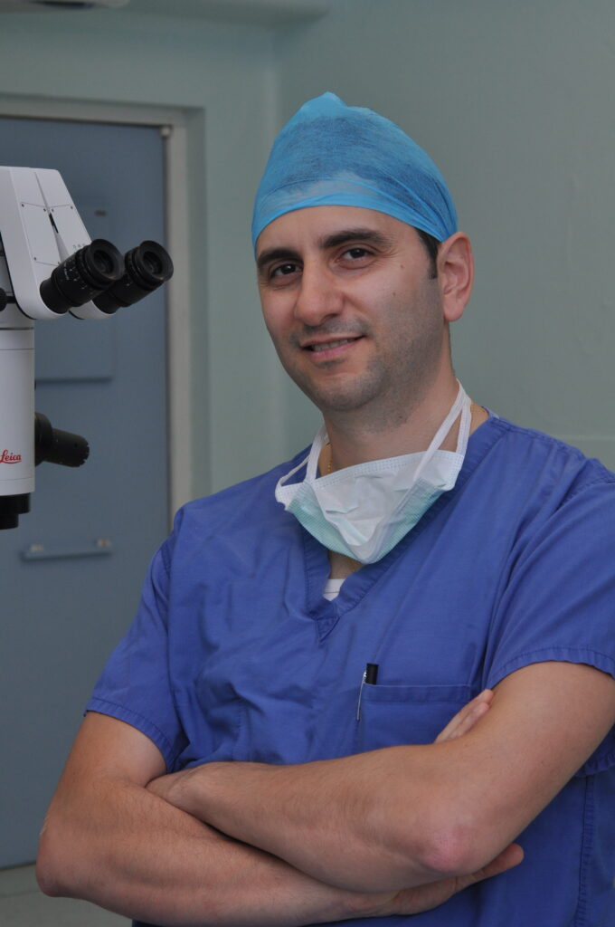
“As part of my routine glaucoma care, I integrate advanced slit lamp imaging to visualise the anterior segment and document angle assessments.
This technology is invaluable for educating patients and ensuring precise monitoring, especially when managing complex cases or following MIGS procedures”.
Mr Chrys Dimitriou, Colchester Primary Eye Care Centre, UK
4. IOP measurements for greater accuracy
Elevated intraocular pressure is the most significant risk factor for glaucoma, making accurate measurement essential. There are two trusted solutions:
Contact Tonometer’s (GATs) – Goldmann Applanation Tonometry for accuracy and precision during slit lamp examinations.
Non-Contact Tonometer’s (NCTs) – Quick, portable screening for routine checks.
Keeler: Your partner in Glaucoma management
Each product in our diagnostic product portfolio is designed to meet the demands of busy eye care practices.
Products suitable for Glaucoma care include:
Slit lamps with exceptional optics and integrated digital capture for documentation and patient education