EyeNews ANTS competition showcases incredible ophthalmic imaging
Keeler
We’re thrilled to share the results of the Eye News ANTS Imaging Competition 2024, proudly sponsored by Keeler, with our Head of Clinical Education, Nicola Bennett, serving as a judge. This competition is a newly established platform aimed at promoting and celebrating the excellence in slit-lamp imaging, offering prizes for three winners and publication in Eye News’ annual imaging supplement.
The response was overwhelming, and the quality of submissions truly speaks for itself. We are proud to recognise the winners, runners-up, and honourable mentions, along with insights from our esteemed panel of judges: Richard Bell, Rosalyn Painter, Nicola Bennett, and Hamza Mussa.
"As one of the competition's judges, 'astonishing' was the first word that sprung to me as I looked through these photographs. It was quite difficult to select not just the winning submission but also the runners-up. There are a lot of things to consider while judging a photography competition, such as composition, exposure, the focusing, meticulousness, and accurate interpretation of the brief."
The winners
First place
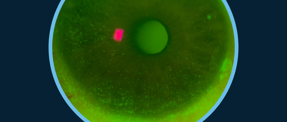
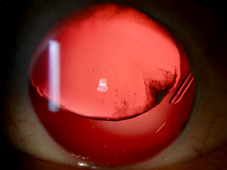
First place - Abigail James, Senior Ophthalmic Photographer, Ophthalmology, Royal Gwent Hospital, Wales, UK.
Abigail’s winning entry captured the intricate details of YAG laser-induced intraocular lens (IOL) damage. This type of damage occurs when the YAG laser, typically used for posterior capsulotomy to treat posterior capsule opacification, inadvertently can impact the IOL material. The resulting photo clearly illustrates damage on the IOL, highlighting the precision required and potential complications of laser treatment.
The judges were particularly impressed by Abigail’s technical approach: she used a thin-to-medium slit beam positioned off-axis, effectively cross-lighting the damage. This technique enhanced the visibility of the fine details and created a sense of depth in the image, allowing for a comprehensive evaluation of the laser-induced changes on the IOL surface.
Second place
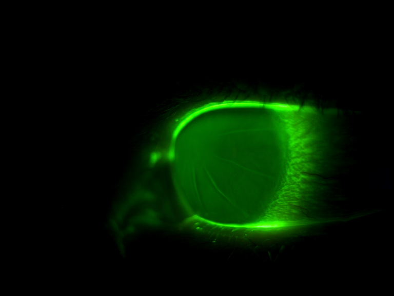
Second place - Keyur Patel, Doctor of Optometry, Tompkins Knight and Son Optometrists, Northampton, UK.
Keyur’s image showcased the corneal flattening that occurs after radial keratotomy (RK), a surgical procedure performed to correct myopia. RK involves making radial incisions in the cornea to change its shape and reduce its refractive power.
The image highlights the post-surgical corneal topography, revealing the distinct flattening effect. Keyur employed sodium fluorescein excitation, which helps illuminate the corneal surface and enhances the visualisation of its contours. He further utilised barrier filters and a diffuser, which improved the clarity and contrast of the fluorescein-stained areas, providing a detailed and vivid representation of the corneal changes induced by the procedure.
Third place
Third place - Vanessa Brebner (Née Shepherd), Senior Medical Photographer Teledermatology, Media Studio, Cambridge University Hospitals NHS Foundation Trust, UK.
Vanessa’s entry utilised retro illumination to showcase aniridia, a rare congenital condition characterised by the complete or partial absence of the iris. Her image vividly captured the associated complications, including the subluxation of the crystalline lens, where the lens is displaced from its normal position, and defects in the zonules, the fibrous structures that hold the lens in place.
By employing retro illumination, Vanessa effectively highlighted the structural abnormalities and the light transmission through the eye, providing a clear and informative view of these complex ocular anomalies. This technique allowed for enhanced visualisation of the lens subluxation and zonule defects, offering valuable clinical insights into aniridia.
Runners up
We also want to highlight two exceptional shortlisted entries.
Shortlist 1 - Robert Hancock
“Iris Flocculus” by Rob Hancock: A striking image of congenital benign iris cysts covering the majority of the pupil.An open slit beam with a wideangle, combined with adjusting the angle ofthe microscope was used to give more depthinformation on the size of the cysts by creatingshadow casting.
Shortlist 2 - Robert Hancock
“Vitreous in the AC” by Rob Hancock: A high-magnification image showing pigment stuck to individual strands of vitreous protruding into the anterior chamber of an aphakic patient.An image taken at x25magnification we can observe pigment stuckto individual strands of the vitreous comingthrough the pupil, into the anterior chamber inthis aphakic patient. Angling the observationsystem off centre allows better visualisation ofthe vitreous protruding anteriorly.
ANTS Imaging Competition 2025
We’re excited to announce that the ANTS Imaging Competition will return next year! Mark your calendars:
Submission window opens: 15 March 2025
Submission deadline: 15 June 2025
Keep an eye on the Eye News website and social media channels for more details as we approach these dates.
At Keeler, we are dedicated to promoting excellence in ophthalmic imaging. The ANTS Imaging Competition has highlighted the remarkable talent and commitment within our ophthalmic community. We sincerely thank all participants, judges, and partners for making this event a tremendous success.
Here’s to advancing the field of ophthalmic imaging and celebrating many more years of the ANTS Imaging Competition!