


The chilling truth about cryotherapy in ophthalmology
Cryotherapy in ophthalmology or ocular cryotherapy explains the application of extremely frigid temperatures to remedy a variety of conditions or disorders concerning the eyes or eyelids. This innovative ophthalmic procedure has been used since the mid-1960s.
Excluding cataract extractions, ocular cryotherapy is typically used as a surface technique — where the probe is applied to the eye or lids without making any incisions into the tissue. Without any incisions, ophthalmic cryosurgery is considered to be much less invasive than other types of ophthalmic incisional procedures or surgeries. Continue reading to learn more about cryotherapy in ophthalmology as well as some of the most common applications.
When is ophthalmic cryosurgery performed and how?
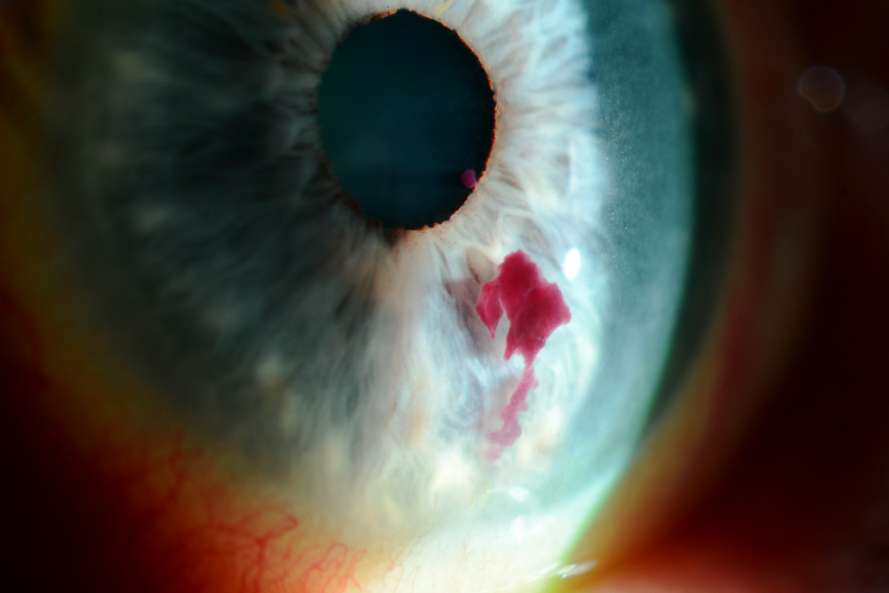
Most often, cryotherapy in ophthalmology is used to treat retinal tears. The process involves freezing the retina surrounding the tear, which creates an adhesive scar around the tear resembling the one created by laser. Ophthalmic cryosurgery is used in a similar manner to when the ophthalmologist performs pneumatic retinopexy and scleral buckling for treating retinal detachments.
Are there ophthalmic cryosurgery advantages or disadvantages over laser treatment?
Considering both methods are designed to accomplish the same result, many ophthalmic surgeons choose cryotherapy out of preference. Both methods take about the same amount of time and offer a similar amount of comfort.
In some instances, however, retinal tears can cause internal eye bleeding, which can block the laser beam from actually reaching the retina. In this instance, cryotherapy is the preferred solution because it freezes from the outside in, which means it’s not impeded by blood on the interior of the eye.
Common applications of cryotherapy in ophthalmology
Applications of cryotherapy in ophthalmology are based on the changes in the tissue caused by temperatures that are below freezing. Upon exposure, living tissue reacts to these sub freezing conditions by forming ice in the extracellular fluid surrounding the cells as well as in the cells.
At the same time, these temperatures cause ice to form inside small blood vessels, which interrupts the supply of blood to adjacent cells. This combination of factors actively destroys living tissue through necrosis and ischemia while inducing inflammation as a reaction to cell death.
In any case, there are several different applications of ophthalmic cryosurgery. Some of the most common applications include cryoextraction of cataracts, retinal cryopexy, cyclocryopexy for advanced glaucoma, and several others.
Cryoextraction of cataracts
The first widely-accepted use of cryotherapy in ophthalmology was cataract surgery. Previously in the 50s and 60s, cataract surgery was conducted by making a 10-12 mm incision with the cataractous lens being totally removed.
Because the lens was grasped with a suction device or capsule forceps, lens breakage was extremely common. In the 60s, an ophthalmic cryoprobe was first used. Shortly after in the 1970s, it became the most common and widely used method for cataract extraction.
Retinal Cryopexy
Retinal cryopexy is commonly used during pneumatic retinopexy and scleral buckling procedures for retinal detachment. In some instances, it’s performed at the time of pars plana vitrectomy to promote chorioretinal adhesions. However, the endolaser technique has largely replaced this practice.
Cyclocryopexy for advanced glaucoma
In the case of advanced intractable glaucoma that can’t be rectified through normal glaucoma surgery or medication, ocular cryopexy has been used to lower the intraocular pressure and reduce aqueous production. This procedure involves the ocular cryopexy being applied to the ciliary body with a transscleral application.
Additional applications of ophthalmic cryosurgery
In addition to the previously-mentioned procedures, ophthalmic cryosurgery is also regularly used to treat:
Discover Keeler ophthalmic cryo surgical products
Keeler has over 40 years experience in offering ophthalmic cryo surgical units. Building on the exceptional reliability of its predecessors, the Cryomatic MKII delivers automation and versatility. The Cryomatic MKII’s coupling system is compatible with a single-use or reusable probe without the need for an adaptor. The cryo console automatically detects the type of probe in use and adjusts internal parameters to reduce the steps required to conduct ophthalmic cryosurgery. The backlit digital display and inbuilt speakers provide the user with intuitive cues such as freeze time, gas cylinder status and probe information.. The Keeler Cryomatic MKII console is compatible with seven reusable probes which are used to treat a variety of pathologies. Keeler also offer a single-use probe reducing the need for sterilisation and improving scalability. Find out more today or get in touch.