ROP screening: The clinical and educational value of paediatric retinal imaging using binocular indirect ophthalmoscopy
Keeler
A focused overview of Retinopathy of Prematurity (ROP), the challenges of paediatric retinal examination, and how shared visualisation support screening, training, and communication.
Understanding Retinopathy of Prematurity (ROP)
Retinopathy of Prematurity (ROP) is a retinal condition affecting premature and low‑birth‑weight infants. It develops because retinal blood vessels, which normally complete maturation in the final weeks of gestation, are still immature at birth. Disruption to this process can lead to abnormal vascular growth and, in severe cases, retinal detachment and permanent vision loss if not identified and managed appropriately.
Why ROP remains a major global concern
ROP remains one of the leading causes of preventable childhood blindness worldwide. Globally, retinopathy of prematurity is estimated to result in over 50,000 cases of childhood blindness or visual impairment each year, highlighting the ongoing need for accurate screening, early detection, and consistent clinical assessment in premature infants (Blencowe etal., 2013; World Health Organization).
ROP is one of the leading causes of preventable childhood blindness worldwide
Infants born before 31 weeks gestation or weighing under 1500g carry the highest risk
With improved neonatal care, more extremely premature infants survive, increasing the number who require careful retinal screening
As neonatal care continues to improve, survival rates among extremely premature infants have increased, resulting in a growing population requiring careful and ongoing retinal screening.
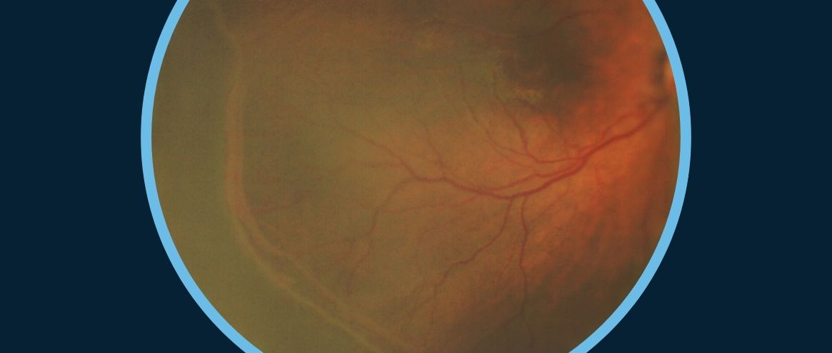
Early ROP changes can be subtle. Vessel dilation, tortuosity, and early ridge formation are often difficult to interpret with confidence, particularly for less experienced examiners. This reinforces the importance of high‑quality visualisation, documentation, and structured training within ROP screening programmes.
Why paediatric retinal examination is particularly challenging
Paediatric retinal examination requires technical skill, clinical judgement, and adaptability.
Clinical challenges include:
Small pupils
Movement or poor co-operation
Sensitivity to light exposure
Need for rapid, yet thorough assessment
Difficulty viewing the far periphery
Subtle vascular changes
As a result, ROP screening relies heavily on examiner experience and tools that support accurate interpretation and communication.
The essential role of binocular indirect ophthalmoscopy (BIO) in ROP screening
Binocular indirect ophthalmoscopy (BIO) is widely recognised as the clinical gold standard for ROP screening and most advances in paediatric retinal imaging are evaluated alongside BIO findings.
BIO enables dynamic, real‑time assessment of the developing retina and offers:
Depth Perception through stereopsis
Wide peripheral view
Ability to Perform indentation
Low‑light options
Flexibility and portability for bedside NICU examination
Peripheral examination is particularly important in ROP, where disease activity often develops outside the posterior pole. Accurate assessment of peripheral vascular changes is central to staging and treatment decisions.
While BIO is essential, it requires experience and structured training. For trainees, learning to interpret subtle findings is especially challenging when they cannot see exactly what the examiner sees during live examinations highlighting the value of shared visualisation tools.
This video shows a ROP examination on a baby, captured using Keeler Vantage Plus Digital
How Video Binocular Indirect tools strengthen clinical training and real‑time assessment:
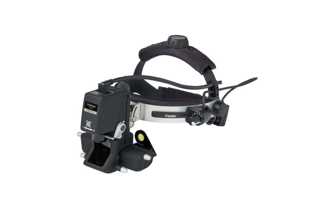
Paediatric devices such as Keeler’s Vantage Plus Digital integrate directly into the BIO workflow. BIO remains the examination method, while VPD enables the examiner’s view including critical peripheral findings to be shared, recorded, and documented.
By transforming an individual examination into a shared visual experience, this supports:
Improved training through real‑time visual guidance
Greater consistency in ROP staging
Clearer communication with NICU teams and families
The complementary role of fluorescein angiography (FA)
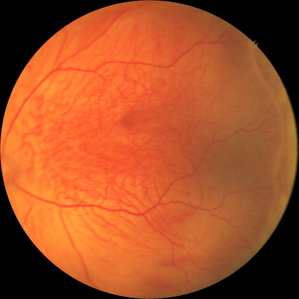
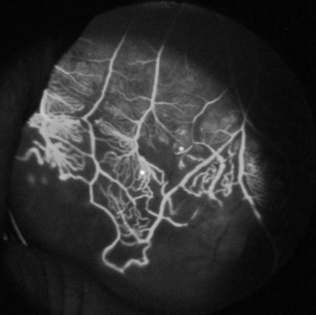
Fluorescein angiography (FA) provides valuable complementary insight whilst using with the BIO, particularly when assessing peripheral retinal vascular behaviour. FA can highlight vascular leakage, avascular retina, and abnormal shunt vessels that may be difficult to appreciate on clinical examination alone.
FA also plays an important educational role, allowing trainees to correlate angiographic findings with clinical BIO observations, strengthening understanding of disease activity and progression. This addition can also enhance parent and patient education by making complex vascular changes easier to explain visually, helping families understand concerns more clearly and feel more engaged in their child’s care.
Final overview
Paediatric retinal examination, especially ROP screening, demands accuracy, confidence, and strong visual interpretation skills. Pairing BIO with VPD technology and FA offers a truly comprehensive approach:
BIO – stereopsis, depth, peripheral view
VPD – real time visualisation, teaching, communication, documentation
FA – enhanced peripheral, visualisation capabilities
Together, these tools support clinicians in delivering high‑quality screening, strengthen examiner training, and improve the consistency of care provided to premature infants.
To learn more about the integration of FA into the indirect workflow using Keeler’s Vantage Plus Digital FA accesory click here
References
Blencowe H, Lawn JE, Vazquez T, Fielder A, Gilbert C.
Preterm‑associated visual impairment and estimates of retinopathy of prematurity at regional and global levels.
The Lancet. 2013;381(9879):2162–2172.
World Health Organization (WHO).
Causes of childhood blindness and visual impairment. WHO reports and global childhood blindness data